
ACMM29
1st - 4th February 2027
EARLY BIRD REGISTRATION CLOSES 15TH NOVEMBER 2026

Workshops

Introduction to Transmission, Scanning Transmission and Analytical Electron Microscopy in Hard and Soft Matter
WSU
Sunday 31st Jan 2027
Presented by: Nestor Zaluzec
In this workshop we will review the basic instrumentation, operating modes and ancillary equipment available in today's modern instruments, as well as discuss imaging (CTEM, STEM, HREM), diffraction(SAED, CBED), and microanalysis (XEDS, EELS) techniques which facilitate characterization from the sub-millimeter to the sub-nanometer scales. The current status of instrumentation, analysis, pitfalls and examples of use as well as considering future prospects will be presented. This workshop will serve as an good introduction to the techniques for the novice as well as a refresher for those who are returning to the field. The application and examples will be taken from both materials and life science perspective.

Advanced FIB for Materials Science
USYD
Friday 5th Feb 2027
Presented by: Dr. Vijay Bhatia/ Dr. Ashalatha Indiradevi/ Dr. Felix Theska/ Dr. Lena Wolff
This workshop is designed for people who already know how to operate a FIB and are looking at advancing their skills. We will be focussing on more complicated techniques such as inverted TEM prep, APT prep, in-situ samples, ion sample interactions, cryo vacuum transfer processes and automation. It will consist of lectures and hands on sessions. There will also be opportunities to network with other FIB experts and share your experience.
The Fine Art of Sample Preparation for Biological Electron Microscopy
USYD
Friday 5th Feb 2027
Presented by: Dr. Errin Johnson/ Dr. Chad Moore
Preparing particulate samples, such as proteins, viruses and vesicles, as well as cells and tissues, for electron microscopy can be a daunting prospect. Good sample preparation is critical to producing high quality data that reflects the native state ultrastructure and processes of biological specimens. There are myriad approaches available, with the most appropriate protocol depending on the sample type and end imaging goal. This workshop is designed to complement the 'Life Science Electron Microscopy Sample Preparation – Past, Present and Future' symposium in ACMM29 and will provide participants with the theory, practical tips and protocols, as well as hands-on experience, for sample preparation of a range of biological samples across all major EM techniques. This includes new stains and approaches for negative staining, TEM and volume EM, as well as cryo-sample preparation and correlative techniques.


Introduction to X-ray Diffraction and Identification of Phases
WSU
Friday 5th Feb 2027
Presented by: Dr. Daniel Fanna and Dr. Ruoming Tian
X-ray Diffraction (XRD) is a valuable complementary technique to SEM and EDS, enabling crystal phase identification and structural analysis of materials. This workshop will introduce the fundamentals of XRD theory and practical operation, covering sample preparation, experimental setup, and basic data analysis including phase identification. Practical tips and troubleshooting strategies will also be discussed, including common artefacts and issues to look out for during data collection and interpretation. The workshop will also provide an overview of micro-diffraction and how it can enhance multimodal materials characterisation workflows.
Fundimentals of Thermal Analysis (STA, TGA, DSC, TMA )
WSU
Sunday 31st Jan 2027
Presented by: Dr. Laurel George and Dr. Andrew Gillian
This workshop introduces the basics of thermal analysis, and how it can be used as a complimentary technique to microscopy and microanalysis for various sample types. Participants will explore the principles of Thermal Gravimetric Analysis (TGA) and Differential Scanning Calorimetry (DSC) and how to identify thermal events such as melting points, thermal degradation and the associated mass loss, phase and glass transitions, crystallisation and more. This workshop will include presentations as well as hands on sample preparation, instrument operation and analysis of real sample data. We will also go through tips and tricks for thermal analysis of different sample types and gas atmospheres.


Advanced Atom Probe Microscopy
USYD
Friday 5th Feb 2027
Presented by: Dr. Andrew Breen/Dr. Kevin Sisco/Leigh Stephenson
Biological and Materials Atomic Force Microscopy
USYD
Friday 5th Feb 2027
Presented by: Dr. Hanyu Li/ Dr. Matt Hadden
Leveraging advances in high-speed and high-sensitivity scanning, Atomic Force Microscopy (AFM) is now essential for nanoscale materials characterisation. This technique precisely measures surface topography, mechanical properties, electrical behaviours, and functional responses.
BioAFM is transforming life science research by opening a window into the nanoscale world of biology. Unlike conventional imaging techniques, it not only delivers breathtaking, high-resolution images of cells, proteins, and biomolecules in their native environments, but also measures their mechanical properties with unmatched precision. This unique combination of visualisation and quantification provides researchers with powerful insights into how biology truly works.
Our workshop provides researchers with a strong theoretical foundation and practical expertise in AFM operation. The program covers core principles and advanced modes, showcasing the instrument's versatility for applications in semiconductors, polymers, 2D materials, and ferroelectrics, cells, tissue and molecules.

AI-driven Single Particle Analysis (SPA) Acquisition for cryo-TEM
UNSW
Sunday 31st Jan 2027
Presented by; Dr. Daniel Luque
This hands-on workshop will introduce AI-driven strategies for single particle analysis acquisition in cryo-TEM, focusing on how automated decision-making can improve data collection efficiency, consistency, and sample targeting. Participants will explore practical workflows that integrate intelligent microscope acquisition with real-time image assessment and downstream processing, enabling faster feedback during data collection and more informed optimisation of acquisition parameters.
Advanced SEM EDS for Low-Z and Air-Sensitive Materials
UNSW
Friday 5th Feb 2027
Presented by: Dr. Raynald Gauvin and Dr. Wu Jiang
This workshop delves into the analysis of samples that challenge conventional SEM-EDS due to weak X ray signals, interaction volume limitations, beam sensitivity, and stringent handling requirements.
Participants will explore:
•Optimized analytical approaches for low Z and nanoscale materials
•Strategies for large area, low-noise high throughput EDS mapping
•Special handling of samples, including preparation methods and inert transfer workflows
This full-day workshop will include a combination of expert talks and live demonstrations on instruments at the UNSW Electron Microscope Unit including the ThermoFisher Scientific Verios FE SEM and CleanMill broad ion beam sample preparation system, and Oxford Instruments Ultim Max Infinity & Unity detectors. Attendees will gain practical insight into how to extend SEM EDS capabilities to materials and scales previously beyond the reach of conventional SEM-EDS.
Attendees will gain practical insight into how to extend SEM EDS capabilities to materials and scales previously beyond the reach of conventional SEM-EDS.

Introduction to 4D STEM: Data Acquisition and Analysis
UNSW
Hilmer Building (E10), lvl5 room:552
Friday 5th Feb 2027
Presented by; Dr. David Muller, Dr. Liam Spillane, Dr. Hiroki Hashiguchi, Dr. Daniel Stroppa
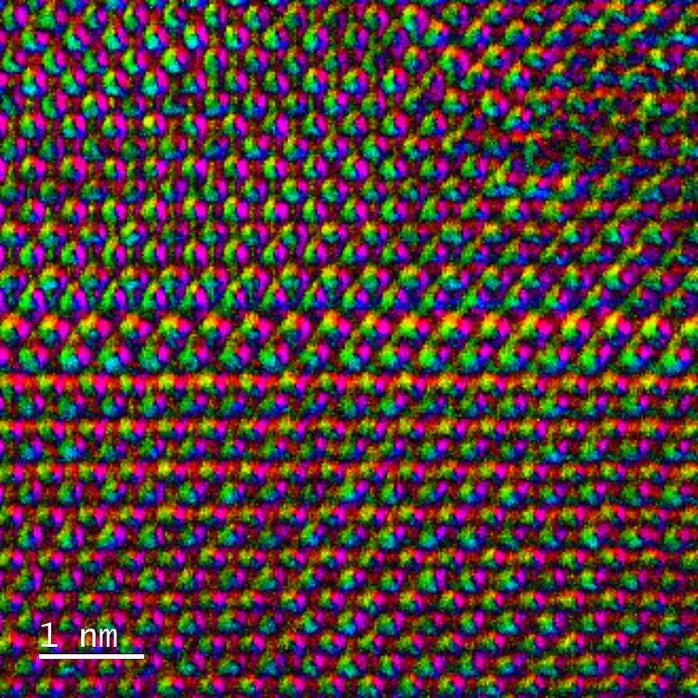
Thanks to recent advances in pixelated direct electron detectors, 4D-STEM (Four-Dimensional Scanning Transmission Electron Microscopy) is enabling new approaches to materials characterisation. By collecting a 2D diffraction pattern for each STEM probe position in a 2D image, we can build a 4D dataset which includes a vast amount of structural and phase information. This lets us map changes in crystal orientation in our samples, stress at grain boundaries and interfaces, electric fields in semiconductors and can be used for low dose and super resolution imaging by capturing ptychographic information. This makes 4D-STEM useful for studying a wide class of materials, including batteries, semiconductors, metals, ceramics, thin films, and other advanced materials used in modern technologies.
The workshop will explore the fundamentals of 4D-STEM also known as scanning electron diffraction (SED) and showcase a range of applications, including:
-
Orientation Mapping
-
Strain Analysis
-
High-Resolution Differential Phase Contrast (DPC)
-
Ptychography
The workshop will begin with introductory talks covering core principles, workflows, and applications for material scientists, followed by demonstrations on data acquisition and analysis on the JEOL GrandARM2 and JEOL F200 TEMs in the EMU at UNSW. Participants will gain practical experience working with real datasets and analysis tools, making this workshop ideal for beginner or expert microscopists looking to expand their characterisation capabilities.
Introduction to Micro-CT: A Practical Guide for Beginners
USYD
Friday 5th Feb 2027
Presented by: Dr. Matt Foley, Dr. Jeremy Shaw
Micro computed tomography (Micro-CT) is a powerful, non-destructive imaging technique that enables three-dimensional visualisation and analysis of the internal structure of materials, biological specimens, and engineered devices at resolution pushing into the sub-micron scale. Widely used across life sciences, materials science, geology, and industrial applications, Micro-CT provides unique insights into morphology, porosity, defects, and complex internal architectures without the need for sectioning or destructive sample preparation.
This small-group, practical workshop is a foundational introduction designed specifically for researchers new to micro-computed tomography. Hosted directly within the SMM Micro-CT facility, the session focuses on the real-world essentials of getting high-quality data while avoiding overly complex physics.
Participants will learn the core basics of sample preparation, scanning geometry, and how to avoid common scanning artifacts. The workshop includes a live demonstration of setting up an acquisition on the scanner, followed by a step-by-step walkthrough of basic 3D data reconstruction and visualisation.
Due to the hands-on nature of the lab environment, this session is strictly capped at 10 attendees.

EM Maintenance
WSU
Friday 5th Feb 2027
Presented by: Dr. Richard Wuhrer
The care and correct maintenance of an SEM is the key aspect for a correctly operating instrument. This workshop will cover many aspects of SEM maintenance. Some of the topics to be covered include: Vacuum Systems and their maintenance, cleaning of microscopes parts such as apertures and wehnelt assembly, basic monitoring of instrument, testing of instrument operation (resolution, magnification calibration), EDS maintenance and calibration, monitoring filament conditions (filament life improvement and setting), and sorting out imaging problems.
Facility Operations Workshop
USYD
Sunday 31st Jan 2027
Presented by: Dr. Paul Macmillan, Dr. Lisa Yen
Being a microscopy facility manager is a complex role, with common challenges faced regardless of the type of platform. This workshop will bring together emerging and current microscopy facility managers from all platform types to discuss strategies and best practices for setting up and running an efficient facility. Through presentations and small group discussions, participants will gain valuable insights and tools to enhance the performance and impact of their facility.
Relevant topics covered will include:
-
How to establish/maintain a microscopy facility
-
Instrument maintenance and Quality Control
-
Researcher training approaches
-
Project onboarding and management
-
Data management approaches
-
Facility staff training & development


Introduction to Expansion Microscopy
UNSW
Friday 5th Feb 2027
Presented by: Dr. Amy Bottomley and Dr. Leeba Ann Chacko
During this hands-on workshop, participants will be introduced to an overview of the principles and practical work-flow of expansion microscopy. The workshop will focus on building both theoretical understanding and practical experience, with emphasis on the key experimental steps required to physically expand biological samples for improved fluorescence imaging.Participants will gain practical experience with key stages of the expansion workflow, including gelation, handling fragile expanded gels, moving and cutting gels, mounting expanded samples, and preparing them for microscopy. The workshop will also cover imaging considerations specific to expanded samples, including gel stability, locating regions of interest, z stack acquisition, and assessing image quality after expansion.The final session will provide an overview of approaches for analysing expansion microscopy data, including key considerations for interpreting expanded samples. Through a combination of talks, demonstrations, and hands on activities, the workshop will give participants a practical introduction to applying expansion microscopy to biological samples.
Fluorescence Lifetime Imaging Microscopy (FLIM)
UNSW
Sunday 31st Jan 2027
Presented by: Dr. Alex Macmillan, Dr. Leeba Chacko, Dr. Taryn Guinan
During this workshop, the Katharina Gaus Light Microscopy Facility will collaborate with Leica Microsystems to highlight the principles and applications associated with fluorescence lifetime–based imaging approaches. The focus will be on building both theoretical understanding and practical skills, including correct experimental setup, lifetime data acquisition, and analysis strategies. Participants will be introduced to methods such as decay fitting and phasor analysis, with applications spanning molecular interactions (e.g. distance measurements using FRET), cellular metabolism (e.g. NADH autofluorescence), and microenvironmental readouts such as pH and ion concentration.
Beyond Conventional FIB: Principle and Applications of Laser-PFIB
UNSW
Sunday 31st Jan 2027
Presented by: Dr. Charlie Kong/ThermoFisher
Ready to push past the limits of traditional Ga-based focused ion beam (FIB) and discover the next frontier of micro-characterisation? Join us for an educational workshop exploring the fundamentals and practical methodologies of emerging FIB technology, featuring multi-gas ion source plasma FIB and cutting-edge femtosecond laser capabilities.
Attendees will explore optimized workflows on 2D/3D applications across diverse research sectors, including advanced materials, additive manufacturing, green energy, next-generation integrated circuits, and multiple engineering disciplines. The group demonstration features a hybrid format, pairing an in-person practical session with a high-fidelity remote livestream of real-time data collection and processing.
Light Sheet Microscopy
UNSW
Sunday 31st Jan 2027
Presented by: Dr. Sandra Fok, Dr. Elvis Pandzic, Dr. Gavin Symonds
This workshop will introduce the fundamentals of light sheet microscopy, a powerful imaging modality for fast, gentle, and high-resolution volumetric imaging of biological specimens. Participants will learn about the optical principles underpinning selective plane illumination and key considerations for sample preparation and experimental design. The workshop will cover both Gaussian and lattice light-sheet modalities, applied to imaging sub-cellular dynamics in live cells, as well as volumetric imaging of organoids and cleared specimens. The session will include practical guidance on data acquisition, optimisation of imaging parameters, and handling large multidimensional datasets. A strong emphasis will be placed on hands-on demonstrations, with participants having the opportunity to directly test the instrumentation. Applications spanning live-cell imaging, organoids, and whole-organism imaging will be highlighted.


Fluorescence Fluctuation Correlation Spectroscopy (FFCS)
UNSW
Friday 5th Feb 2027
Presented by: Dr. Elvis Pandzic, Dr. Alex MacMillan, Dr. Gavin Symonds, Dr. Liz Hinde
This workshop provides an introduction to a suite of light microscopy techniques for quantifying molecular density, dynamics, and co-localization. Through a combination of theoretical discussion and practical demonstrations, participants will explore how different acquisition modalities—including spot scanning, line scanning, and image series—within fluorescence fluctuation correlation spectroscopy can be leveraged to enhance temporal and spatial resolution, as well as increase information content.
Session details:
The morning session will begin with an introductory lecture outlining the principles behind these modalities and their application to quantifying parameters such as molecular concentration, diffusion coefficients, velocities, kinetics, and interactions. This will be followed by a practical session focused on the simulation of microscopy image data and their analysis using FFCS tools. In the afternoon, participants will engage in hands-on sessions, gaining direct experience with these techniques on both laser scanning confocal and camera-based systems.
Fiji and Friends - AI and ML Image Analysis with Open Source Software
MQ
Friday 5th Feb 2027
Presented by: Dr. Cameron Nowell
This workshop will be presented in a hands-on, masterclass type format. Users will be given various imaging data sets to work through. Initial data will be analysed using canned AI models in CellPose with machine learning from lastik. Later data will have users learning how to train their own AI and ML models and apply these in automated code to batch analyse image sets.
Requirements: You must bring your own laptop, with charger and an external mouse (tracing things with a touchpad is not fun). Before the workshop, you will be expected to install and configure open source software (details and instructions will be provided to attendees). Please note that the installation instructions have only been tested on Windows, not Mac. Mac install follows similar steps but can hit some issues.


Quantitative X-Ray Microanalysis with Monte Carlo Simulations
WSU
Sunday 31st Jan 2027
Presented by: Dr. Raynald Gauvin
This full-day workshop will cover the fundamentals and limitations of quantitative X-ray Microanalysis in the TEM and in the SEM for thin films and bulk specimens. The main limitation of quantitative x-ray microanalysis is that specimens must be homogeneous in composition and have well-defined geometry: parallel surfaces for thin films and flat surfaces normal to the electron beam for homogeneous materials. It will be shown how Monte Carlo simulations can quantify more complicated geometries and non-homogeneous materials with the well-known software Casino and MCX-Ray. The participants are invited to bring their laptops since software will be provided and live demos will be performed.
Micro Electron Diffraction (MicroED)
UNSW
Friday 5th Feb 2027
Presented by: Dr. Hongyi Xu, Dr. Sulin Li, Dr. Felipe Kremer
MicroED is a rapidly emerging technique that provides an alternative to conventional single crystal x-ray diffraction (XRD). Owing to their higher scattering probability, electrons allow for the study of specimens which are otherwise untreatable by traditional XRD (i.e samples in the nanometre size range). With applications spanning structural biology, pharmaceutical research, chemistry, and materials science, MicroED is becoming an essential tool for structural characterisation of both biological and non-biological specimens.
This workshop will provide a practical introduction to MicroED workflows, covering the theoretical background of electron diffraction and the principles underlying MicroED. Attendees will learn about sample preparation strategies, methods for 3D electron diffraction (3D ED) data collection*, and approaches for 3D ED data processing. The workshop will also explore structure solution and refinement pathways for small molecules and proteins**. Participants will gain an understanding of the capabilities, challenges, and applications of MicroED, enabling them to evaluate and implement this powerful technique in their own research programs.
* Remote demonstration session.
** Hands-on session. Attendees are recommended to bring their own computers.


Unravelling the Nanoarchitecture of Life: Breakthroughs with Time-Resolved STED Nanoscopy
USYD
Friday 5th Feb 2027
Presented by: Dr. Neftali Flores Rodriguez and Dr. Shelly Azadi
The Power of Time: Leveraging Tau-STED and FLIM Mechanics
Moving beyond raw laser power. How lifetime-based separation and time-gating drastically improve spatial resolution while minimising phototoxicity and photobleaching on delicate samples.From Sample to Publication: Optimising the STED Pipeline the critical choice of fluorophores (matching depletion lines), depletion-hardy mounting media, and labelling density. How to design an experiment that succeeds before it even hits the stage. Resolving Complex Biology: Multi-Color and Dynamic NanoscopyReal-world applications: Resolving structural biology, organelle contact sites, and molecular clustering at the nanoscale in both fixed and living systems.
An Introduction to Infrared and Raman Microscopy for Mapping and Imaging
USYD
Friday 5th Feb 2027
Presented by: Der. Liz Carter/ Dr. Michelle Wood
This workshop introduces infrared (IR) and Raman spectroscopy, showcasing the capabilities of modern systems for chemical mapping, imaging, and micro-scale analysis. Participants will learn how these non-destructive techniques can characterise materials at micron and even sub-micron scales with ease and precision. Using examples from material sciences, life sciences, and cultural heritage, this workshop will demonstrate how advanced mapping and imaging techniques enable the visualisation of the spatial distribution of components within a sample, including proteins, lipids, and carbohydrates in biological tissues, or various pigments and binders in historical artworks. These techniques allow for detailed analysis without the need for external markers or tagging. In addition to compositional mapping, the workshop will highlight how these methods can be used to investigate changes that occur due to stress and strain, offering insights into mechanical and structural transformations. This session is ideal for those new to the field, offering an accessible starting point, as well as for experienced users seeking a refresher or update on recent instrumentation developments.
